

The Mechanical Stage Upper Knob
Microscope Stages
All microscopes are designed to include a stage where the specimen (usually mounted onto a glass slide) is placed for observation. Stages are often equipped with a mechanical device that holds the specimen slide in place and can smoothly translate the slide back and forth as well as from side to side. A stage can be classified according to design and functionality. In the simplest case, the plain phase (illustrated in Figure 2, on the left) consists of a rectangular or square design containing several clips to concord the specimen slide.
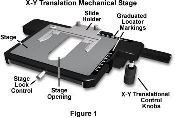
The stage illustrated in Figure one is a typical rectangular stage with the added feature of a specimen slide translational control device normally referred to as a mechanical phase. This mechanical stage contains controls for right-handed microscopists that allow the movement of the specimen slide in both the X (correct and left) and Y (back and along) directions so the microscopist can examine the unabridged microscope slide (secured to the phase with the slide holder). Similar mechanical stages are available with the translational control knobs situated on the left-manus side of the phase for left-handed microscopists. The stage illustrated in Figure one also contains a rather large opening in the center to allow light from the condenser to pass through the specimen (the stage opening). The stage is also equipped with a locking control that allows the phase to be stock-still into position with respect to its rotation around the condenser axis. Graduated locator marks positioned on the mechanical portion of the stage allow the microscopist to note the location of important specimen details, assuasive piece of cake render to the area for additional ascertainment or photomicrography.
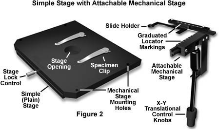
A unproblematic (commonly termed "plainly") microscope stage is illustrated on the left in Figure 2. This stage contains an opening to admit light from the condenser, several mounting holes for a mechanical phase, and two clips that secure the specimen slide in identify for observation under increasing magnification (changing of objectives) and for photomicrography. The stage is besides equipped with a locking control that can prepare the position of the stage. This stage is very useful for quick examination of specimens, merely is very hard to use with higher power objectives (above 20X). At high magnification, small-scale translations of the specimen slide will usually throw the features of interest completely out of the viewfield, and attempting to relocate them can lead to frustration. Auxiliary mechanical stages attached to a simple stage can allow for minute translation of the specimen slide, making it easier for the microscopist to detect specific areas on the slide. The mechanical stage attachment illustrated on the right in Figure 2 can be hands attached to the unproblematic stage on the left in the same figure. In order to attach this mechanical stage, the specimen clips must beginning be removed.
Note that the stage opening is much smaller on the stage illustrated in Figure two when compared to the mechanical phase shown in Effigy i. This is considering the mechanical portion of the stage in Figure 1 encompasses the upper stage plate allowing the greyness role of the stage to move with the specimen slide holder in both the 10 and Y directions. Alternatively, when an auxiliary mechanical stage (like to the one on the right in Figure 2) is attached to the simple stage shown in Figure two, it is only the mechanical slide holder that moves, and the stage itself remains stationary. Locator graduations on the attachable mechanical stage (that help identify specific areas of a specimen) operate in a manner like to the mechanical phase illustrated in Effigy i. Both stages may be rotated with respect to the microscope torso to facilitate framing during photomicrography. The stages illustrated in Figures 1 and two are not centerable, but many designs (including some discussed beneath) are capable of being centered with respect to the optical axis of the microscope.
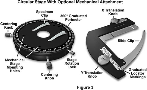
The circular graduated stage, illustrated on the left in Effigy 3, is one of the nearly versatile and useful designs for all types of microscopy and photomicrography. These stages rotate 360�, permitting complete rotation of the samples and bang-up ease in fine-tuning the limerick of viewfields for photomicrography. An added feature is the graduations included on the periphery of the stage, which allow for precise alignment when critical belittling measurements must be undertaken. The phase rotates on brawl bearings that provide precision rotation without annoying jerks, bumps, and stalls. 2 centering knobs (Figure 3) permit the stage to exist centered with respect to the optical centrality of the microscope. When used with objectives in a centering nosepiece, the microscope tin be adjusted to be parcentric, significant that a specimen centered in the field of view for one objective remains centered when the nosepiece is rotated to bring some other objective into employ.
An optional mechanical stage zipper (on the right in Figure iii) for the circular stage permits accurate translation of samples like to the mechanical stages discussed in a higher place. About circular stages have pre-drilled mounting holes that let positioning and firm zipper of the optional mechanical stage. Circular stages also have a locking mechanism (stage rotation lock) that let them to exist locked into a single position. These stages often are equipped with click stops that alert the microscopist at 45� intervals of rotation.
Mod microscope stages are precisely manufactured and very durable. They are cast from either iron or aluminum and automobile-finished to accurate tolerances. Translation mechanisms are usually rack-and-pinion styles, machined from aluminum, brass, or (more recently) synthetic polymers. The synthetic gear mechanisms normally are far less durable than their metal counterparts, especially afterward years of use. These devices should be thoroughly cleaned, lubricated, and tightened for adjustment every several years. Most stages are protected with a very tough non-glossy ceramic coating that resists marks and scratches. The dull surface ensures that stray calorie-free is non reflected from the stage into the objective.
Specialized Microscope Stages - In that location are a wide variety of microscope stages that are designed for specific purposes. These include stages equipped with auxiliary equipment for manipulating samples during ascertainment, measuring systems that permit precise measurements to exist made over very small distances, universal stages that allow measurements over many specimen angles, and other specialized stages that perform a myriad of unique functions. We will hash out some of the more than common specialized microscope stages below.
Inverted Microscope Stages - Inverted microscopes are configured differently than the standard upright microscope. These microscopes have the objectives placed below the phase and use several different condenser configurations to illuminate the specimen. Tissue civilization microscopes have a condenser that is mounted higher up the stage, while epi-illumination microscopes (metallographs and their relatives) have a substage condenser that preceeds the objectives. In both cases, the stage is slightly modified from standard stages as illustrated in Figure 4 beneath.
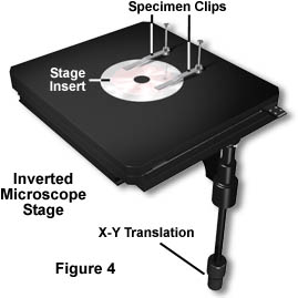
The inverted microscope phase is similar in its basic overall design to the mechanical stage illustrated in Figure 1. Both stages have translational controls that allow the phase (and specimen) to be moved in both the Ten and Y directions. The master divergence is the large phase opening that accommodates an insert on the inverted microscope phase. The inserts (Effigy four) are ordinarily fabricated of stainless steel and have openings of various sizes to let for big differences in sample size. For instance, with inverted tissue culture microscopes, researchers oftentimes must scan large culture flasks to observe the entire population of cells and this is much easier to accomplish when the stage insert has a large opening. Metallography samples are likewise often quite large and more of their surface can be observed with the specialized inserts. When minor specimens are to be observed, an insert with a very minor opening (near the size of the one in Figure 4) is used to support the sample for observation. Inverted microscope stages do not translate up and down. Focusing is achieved using a translatable nosepiece that, together with the objectives, moves up and down.
High-Magnification Measuring Systems - Quantitative microscopy often demands highly accurate measurements of diverse specimen dimensions. These include measurement of miniature precision parts and semiconductors as well equally high-precision assembly of magnetic heads and other minute electronic components. Specialized microscopes with precision stages are often used for these purposes. The stage illustrated in Figure 5 is an instance of a precision microsope stage. It is equipped with a micrometer-fashion translational apparatus that allows for very accurate and controlled movements of the phase and specimen. The phase is also rotatable over the unabridged 360� range for total control over the measurements.
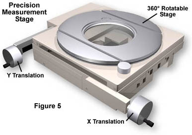
Microscopes designed for stages of this type are sometimes equipped with prisms for Cock Epitome Ascertainment, meaning that the observed image and the specimen both move in the same direction (the contrary is true for virtually microscopes). The stages are commonly accompanied by electronic controllers that requite authentic digital readouts of the stage position, and most tin exist programmed for precise stage movements in a sequence of steps. There are a wide multifariousness of designs for these specialized stages and many aftermarket manufacturers are capable of supplying custom-configured stages built to the client's specifications.
Micromanipulators - It is often necessary to manipulate the specimen while it is beingness observed under the microscope. This is the case in many tissue civilization and in vitro fertilization experiments besides as genetic implantation procedures that crave shut observation of the sample during the experiment. The micromanipulator stage illustrated in Figure 6 is an case of the type of equipment necessary to behave these types of operations.
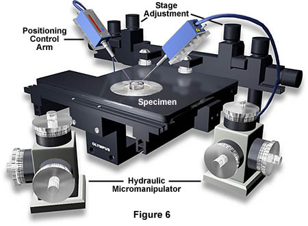
This specialized phase has two triple-axis hydraulically controlled water micromanipulators that orchestrate specimen handling in a highly accurate manner. In the analogy, a modest petri dish containing embryos is being manipulated with the control arms to monitor pH of the solution besides as providing radioactively labeled nutrients to the embryos. These manipulators can be adjusted to perform a broad spectrum of functions ranging from microinjection to electrochemistry experiments. There are a number of manufacturers who supply micromanipulators and accessories that adhere to commercial microscopes such as those manufactured past Olympus and Nikon.
Universal Phase - This unwieldly looking microscope stage permits tilting of a thin specimen at whatever bending for measuring the optical structure of a birefringent crystal. The universal phase illustrated in Figure 7 is an instance of this type of phase.
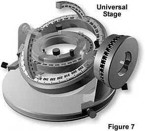
Universal stages are designed to be used with special long working distance (LWD) objectives and very low magnifications usually ranging from 5X to 20X. These stages are graduated on all 4 axes with rotation scales that are distinguished by different color codes. The four rotating centers of the main trunk all rest on a common betoken. Two hemispherical lenses are used to sandwich the specimen between their plane surfaces with immersion oil being applied to all contact surfaces.
Universal Stage Objective Specifications
Table ane
The universal stage attaches to the circular petrography stage illustrated in Figure iii using screws permanently fixed onto the universal stage. There is a primal ring on the microscope stage that must start exist removed before installation and centering of the universal stage. The stage contains locking devices that permit the position of the sample to be fixed for viewing and photomicrography. Universal stages are no longer being manufactured past the major microscope producers, but a limited number are however available through distributors and custom aftermarket manufacturers.
There are a variety of other specialized microscope stages that we have not covered in this give-and-take. Rapid growth in the semiconductor arena has led to the design and manufacture of a number of stages utilized to examine and dispense integrated circuit wafers. Similar microscopes have also been adapted in other areas of integrated circuit manufacture. Many scientists design and build their own custom stages for specific experiments including biomedical investigations, cell manipulation, materials research, and geology.
Contributing Authors
Mortimer Abramowitz - Olympus America, Inc., Two Corporate Center Drive., Melville, New York, 11747.
Michael W. Davidson - National High Magnetic Field Laboratory, 1800 East Paul Dirac Dr., The Florida State University, Tallahassee, Florida, 32310.
BACK TO ANATOMY OF THE MICROSCOPE
Questions or comments? Send us an email.
© 1998-2022 by Michael West. Davidson and The Florida State Academy. All Rights Reserved. No images, graphics, scripts, or applets may be reproduced or used in whatever way without permission from the copyright holders. Use of this website means you concur to all of the Legal Terms and Conditions prepare along by the owners.
This website is maintained by our
Graphics & Spider web Programming Team
in collaboration with Optical Microscopy at the
National High Magnetic Field Laboratory.
Last modification: Friday, November thirteen, 2015 at 02:xviii PM
Admission Count Since September 6, 1998: 125951
For more information on microscope manufacturers,
use the buttons below to navigate to their websites:

The Mechanical Stage Upper Knob,
Source: https://micro.magnet.fsu.edu/primer/anatomy/stage.html
Posted by: stewartantim1964.blogspot.com
0 Response to "The Mechanical Stage Upper Knob"
Post a Comment